Unitma Digital Histology
Product Catalogue
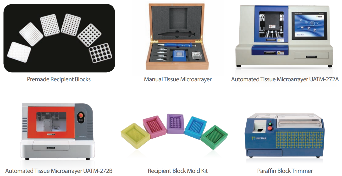
* Premade Recipient Block
* Manual Tissue Microarrayer
* Automated Tissue Microarrayers
* Recipient Block Mold Kit
* Paraffin Block Trimmer
Tissue Microarray (TMA)
Tissue Microarrays are a collection of multiple tissue cores that are arranged in columns and rows inside a paraffin
block allowing for histological analysis. They are a crucial tool in the analysis of gene and protein expression levels in
samples from normal and diseased specimens. Further, they are useful in the early-stage discovery of gene targets
in genomic research, validating targets, testing, and optimization of diagnostic tests, and in the quality control of
molecular detection schemes.
And the TMA technology not only makes decrease the reagent, time, and human resource below one sixtieth but
also can be applied to most of the know-how about tissues for immunohistochemistry, in situ hybridization, FISH
and in situ PCR.
TMA advantages
• The use of relatively small tissue cores to be positioned onto a unique slide allows simultaneous histological analysis
of hundreds of samples.
• High throughput screening of expression
• Reduction of the number of slides to be mounted and analyzed → Time saving
• Reduction of the amount of reagents and antibodies used → Money saving
• Analysis of various biological specimens under uniform reaction conditions → Increasing the consistency of the
results
• Saving of precious biological samples and possibility to re-use donor blocks
UNITMA TMA technique
In conventional Tissue Microarray techniques, all the users have to prepare for their recipient blocks by drilling the
dummy paraffin blocks manually prior to starting TMA work.
But Unitma developed the premade paraffin recipient blocks for the first time in the world. The premade recipient
blocks have the well rounded hole matrix suitable for the digital histologic analysis.
Furthermore, Unitma’s premade recipient block brings to save the time and cost in preparation of the desired
recipient blocks manually and high throughput with a higher TMA success rate up to 99%.
Based on the core technology, Unitma developed the manual and automated tissue microarrayers and has been
supplying the most innovative instruments with the premade recipient blocks to worldwide customers as the best
solution for the digital histology.Recently Unitma also launched the automated paraffin block trimmer which can
trim the paraffin residues around the paraffin embedded tissue blocks without any damages to the sample tissues
and any injuries during the trimming work by the conventional trimming way. This instrument is only a paraffin block
trimmer without any competitive instruments in the current world market.Today, Unitma is continuously developing
new technologies to change the existing histologic techniques.
Premade Recipient Blocks
Automated Tissue Microarrayer UATM-272B
Manual Tissue Microarrayer
Recipient Block Mold Kit
Automated Tissue Microarrayer UATM-272A
Paraffin Block Trimmer
02 03
Certification Premade Recipient Block &
Recipient Block Builder
Manual Tissue Microarrayer
Automated Tissue Microarrayer
Paraffin Block Trimmer
Premade Recipient Blocks
Unitma provides the premade recipient blocks
patented in global to save the valuable time and cost
in creating the recipient blocks additionally before
starting TMA work.
The premade recipient block is made of special
material which melts when heated at about 70 °C for
30~60minutes.
The blocks have evenly spaced round wells arranged
in a square matrix which conforms to digital
pathology trend.
Currently, six different sized blocks are available
0.5mm (320 holes), 1mm (120 holes), 1.5mm (90
holes), 2mm (60 holes), 3mm (30 holes), 5mm (20
holes)
The blocks of 1, 1.5, 2, 3, 5mm applicable to manual
Tissue Microarrayer while the blocks of 0.5, 1, 1.5,
2mm applicable to Automated Tissue Microarrayer
(UATM-272B)
Features
• TMA success rate up to 99%
• Maintains tissue integrity during embedding
• Up to 300 tissue slices per a block
• High durability during microarraying
• Standardized block configuration for the simple
image analysis
• Saving the additional time and cost in preparation of
recipient blocks
• Well rounded hole matrix suitable for digital
pathology
• Ready-to-use paraffin recipient block
• Compact design and versatility
• No dedicated space required for TMA block
formation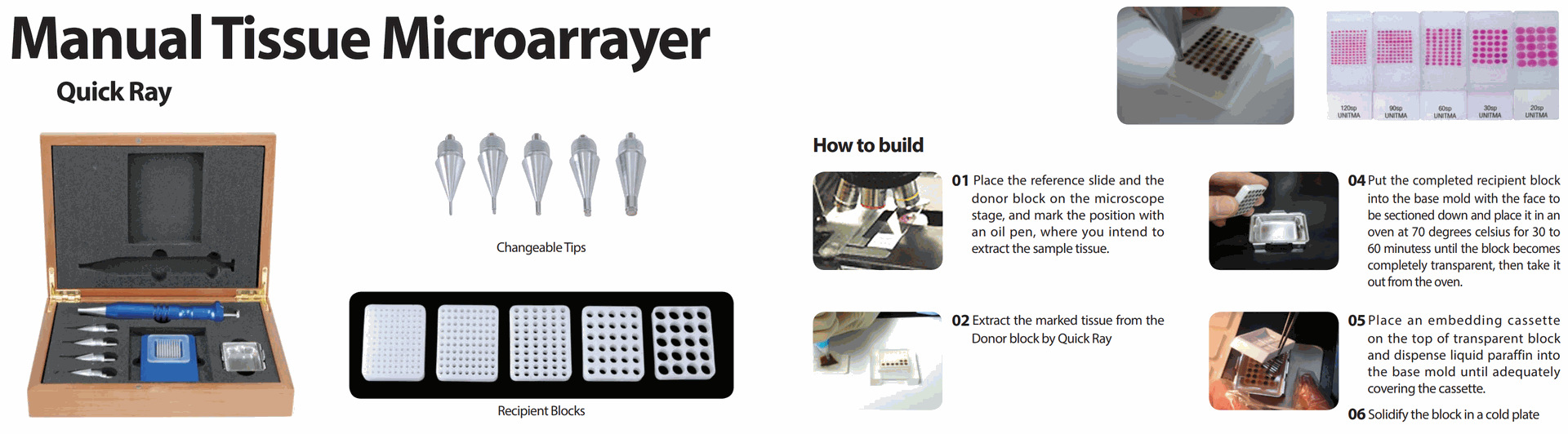
In Situ Hybridization
0.5mm(320 holes) 1mm(120 holes) 1.5mm(90 holes) 2mm(60 holes) 3mm(30 holes) 5mm(20 holes)
FISH Special stain Immunohistochemistry
Product name Premade Recipient Block
Model Number Core size Weight Number of cores
UB06-0.5 0.5 mm 3.4g 320 (16 x 20) holes
UB06-1 1 mm 3.2g 120 (10 x 12) holes
UB06-1.5 1.5 mm 2.8g 90 (9 x 10) holes
UB06-2 2 mm 2.5g 60 (6 x 10) holes
UB06-3 3 mm 2.5g 30 (5 x 6) holes
UB06-5 5 mm 1.8g 20 (4 x 5) holes
Dimension 24 x 30 x 5.5 mm
Color and odor White, odorless
Physical state and appearance Solid
Storage temperature Room temperature
Operating temparature 5°C ~35°C
Composition Paraffin plus special materials
Purpose of use Research purpose only
Certification ISO, CE
Manual Tissue Microarrayer
The newest technique for superior
quality tissue microarrays (TMAs)
The Quick Ray system represents the newest technique
available for producing superior quality Tissue Microarrays
(TMAs) in less time than with traditional methods at a
fraction of the cost.
The Quick Ray technique produces superior quality
TMAs in less time than with traditional methods, and
accomplishes finished products at a fraction of the cost.
The Quick Ray uses a hollowed tip to remove tissue
cores as small as 1mm from paraffin-embedded tissue
(donor block). The extracted tissue is then inserted into
a preformed recipient block-the Quick Ray System is
the only system available that uses a preformed paraffin
recipient block.
The finished block is sectioned using a microtome; the
sections are mounted on glass slides and then stained.
Each TMA block can be cut into 300~400 sections at
4~5μ, which can then be subjected to independent
tests. Common tests include immunohistochemistry and
fluorescent in situ hybridization. Compact design and
versatility make building a Quick Ray TMA astoundingly
simple. The construction of a TMA block can be done on
any clean counter, and does not require a dedicated space.
Features
• Portable and easy to handle
• Shortening the TMA work
• Smarter arrayer compared to conventional products
• Easy to carry and to make the array block anytime &
anywhere
• Inexperienced pathologist can be easily familiar with
the kit
• Simple procedure for creating the blocks
• Easy to create the various sized blocks by using the
premade recipient blocks
• Save the time in preparation of the recipient blocks
Recipient Blocks
Changeable Tips
01 Place the reference slide and the
donor block on the microscope
stage, and mark the position with
an oil pen, where you intend to
extract the sample tissue.
04Put the completed recipient block
into the base mold with the face to
be sectioned down and place it in an
oven at 70 degrees celsius for 30 to
60 minutess until the block becomes
completely transparent, then take it
out from the oven.
02 Extract the marked tissue from the
Donor block by Quick Ray
05Place an embedding cassette
on the top of transparent block
and dispense liquid paraffin into
the base mold until adequately
covering the cassette.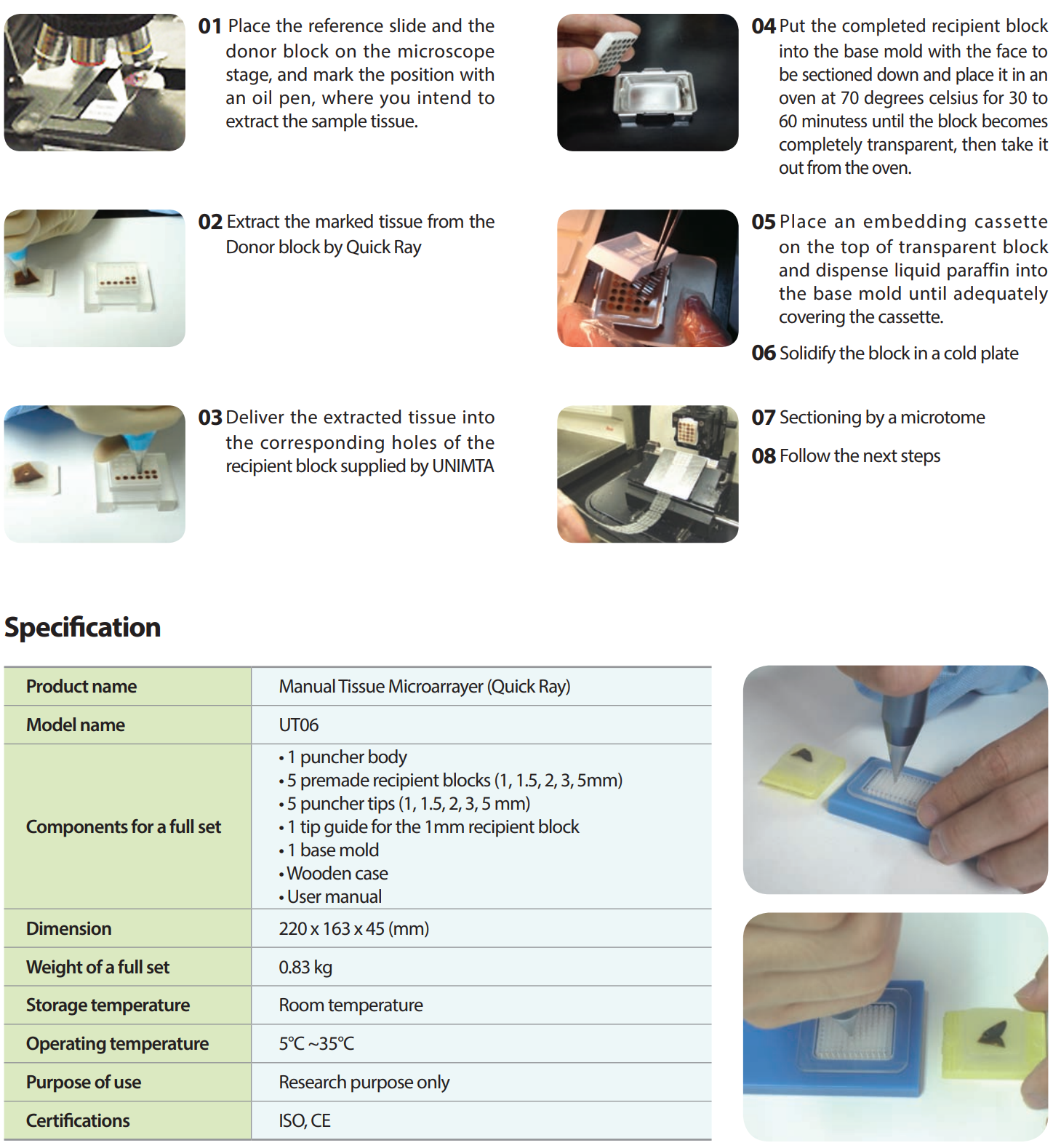
06 Solidify the block in a cold plate
03Deliver the extracted tissue into
the corresponding holes of the
recipient block supplied by UNIMTA
07 Sectioning by a microtome
08 Follow the next steps
How to build
Specification
Product name Manual Tissue Microarrayer (Quick Ray)
Model name UT06
Components for a full set
• 1 puncher body
• 5 premade recipient blocks (1, 1.5, 2, 3, 5mm)
• 5 puncher tips (1, 1.5, 2, 3, 5 mm)
• 1 tip guide for the 1mm recipient block
• 1 base mold
• Wooden case
• User manual
Dimension 220 x 163 x 45 (mm)
Weight of a full set 0.83 kg
Storage temperature Room temperature
Operating temperature 5°C ~35°C
Purpose of use Research purpose only
Automated Tissue Microarrayer
Quick Ray Master is an automated tissue microarrayer
running by the built-in PC. 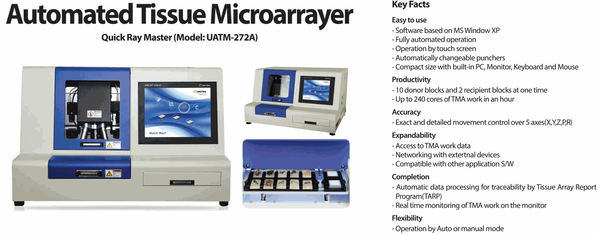
The instrument extracts the sample tissues from the donor
blocks and delivers the extracted sample tissues into
the correspondent hole of the premade recipient block
automatically.
And the researcher can access to the data file for the
current TMA work by using the Tissue Array Report
Program (TARP) developed by UNITMA.
The researchers can choose one of the 3 rotary type tips
simply by using unique UNITMA software. The instrument
can be connected to the external devices such as USB,
monitors, printers and etc.
The instrument provides economical efficiency in time,
quality, and cost of tissue preparation by fully automating
tissue microarraying process.
Features
• Analysis of many patient tissue samples simultaneously
• Conserves tumors, biopses and other precious
biological samples
• Conserves antibodies and other expensive reagents
• Improves assay precision through sample and
patient multi-plexing
• Applicable to mRNA and protein expression analysis
• Miniaturize and automate immunohistochemistry, in
situ hybridization, FISH, and in situ PCR
• High-speed preparation of tissue microarray blocks
• Maintains tissue integrity during embedding
• Up to 300 tissues slices per recipient block
• Standardized block configuration simply image analysis
• Prepare up to 240 tissue cores per hour
• TMA success rate up to 99%
• Tissue array circle time 15secs
• Positional accuracy of 5μm at recipient blocks
Key Facts
Specification
List Description
Product name Quick Ray Master (Model : UATM-272A)
Instrument type Automated Tissue Microarrayer
Punch type Rotary puncher(1mm, 2mm, 3mm)
Power supply 100 to 120 VAC, 200 to 240 VAC / 50~60Hz
Space required 953 x 703 x 610mm (W x D x H)
Weight 148kg
Stage capacity 10 donor blocks and 2 recipient blocks
Speed 1 cycle time : 15 seconds (Pick & Place)
Monitor LCD monitor 15"(1024x768) with a touch screen
Camera resolution Camera 1 : 1280 x 1024, Camera 2 : 2,592 x 1,944 (pixels)
Operating system Unique UNITMA S/W by MS Window XP
Recipient block Premade recipient block to be supplied by UNITMA
Certifications ISO, CE, FCC, UL
Quick Ray Master (Model: UATM-272A)
Easy to use
- Software based on MS Window XP
- Fully automated operation
- Operation by touch screen
- Automatically changeable punchers
- Compact size with built-in PC, Monitor, Keyboard and Mouse
Productivity
- 10 donor blocks and 2 recipient blocks at one time
- Up to 240 cores of TMA work in an hour
Accuracy
- Exact and detailed movement control over 5 axes(X,Y,Z,P,R)
Expandability
- Access to TMA work data
- Networking with extertnal devices
- Compatible with other application S/W
Completion
- Automatic data processing for traceability by Tissue Array Report
Program(TARP)
- Real time monitoring of TMA work on the monitor
Flexibility
- Operation by Auto or manual mode
Automated Tissue Microarrayer Key Facts TARP file
Specification
Product name Quick Ray Master (Model : UATM-272B)
Instrument type Automated Tissue Microarrayer
Tip type Changeable Tips (0.5mm, 1mm, 1.5mm, 2mm)
Power supply 220 VAC, 50/60Hz
Dimension (W x D x H) 750 x 600 x 600 (mm)
Weight 80kg
Capacity 10 donor blocks and 2 recipient blocks
Speed 1 cycle time : 10 seconds
Camera resolution 2,592 x 1944 (pixels)
Operating system Unique UNITMA S/W by Window 7
Recipient block Unitma’s premade recipient blocks
Certifications ISO, CE, FCC, UL
Quick Ray Master (Model: UATM-272B)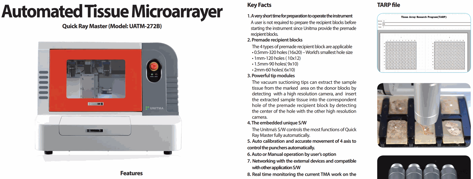
1. A very short time for preparation to operate the instrument
A user is not required to prepare the recipient blocks before
starting the instrument since Unitma provide the premade
recipient blocks.
2. Premade recipient blocks
The 4 types of premade recipient block are applicable
• 0.5mm-320 holes (16x20) – World’s smallest hole size
• 1mm-120 holes ( 10x12)
• 1.5mm-90 holes( 9x10)
• 2mm-60 holes( 6x10)
3. Powerful tip modules
The vacuum suctioning tips can extract the sample
tissue from the marked area on the donor blocks by
detecting with a high resolution camera, and insert
the extracted sample tissue into the correspondent
hole of the premade recipient block by detecting
the center of the hole with the other high resolution
camera.
4. The embedded unique S/W
The Unitma’s S/W controls the most functions of Quick
Ray Master fully automatically.
5. Auto calibration and accurate movement of 4 axis to
control the punchers automatically.
6. Auto or Manual operation by user’s option
7. Networking with the external devices and compatible
with other application S/W
8. Real time monitoring the current TMA work on the
monitor
9. High productivity up to 240 holes per hour
10. Not needed the measurement of the donor block
height
11. Smarter and easier User Interface to run the instrument.
12. Easy TMA data management by Excel data sheet
Quick Ray Master is an automated tissue microarrayer
running by simply connecting to user’s computer
through USB port. The system extracts the sample
tissues from the donor blocks and delivers the
extracted sample tissues into the correspondent hole
of the premade recipient block automatically. And the
researcher can access the data file for the current TMA
work by using the Tissue Array Report Program (TARP)
developed by UNITMA.
The 4 different sized tips are manually changeable to
associate with the desired core size of the recipient
blocks: 0.5mm(320 holes), 1mm(120 holes), 1.5mm(90
holes), 2mm(60 holes). UATM-272B is smarter
instrument compared to UATM-272A, and is designed
for the users to be easily familiar with.
The instrument provides economical efficiency in
time, quality, and cost of tissue preparation by fully
automating tissue microarraying process.
Features
• Analysis of many patient tissue samples simultaneously
• Conserves tumors, biopses and other precious
biological samples
• Conserves antibodies and other expensive reagents
• Improves array precision through sample and patient
multi-plexing
• Applicable to mRNA and protein expression analysis
• Miniaturize and automate immunohistochemistry, in
situ hybridization, FISH, and in situ PCR
• High-speed preparation of tissue microarray blocks
• Maintains tissue integrity during embedding
• Up to 300 tissues slices per recipient block
• Standardized block configuration simply image analysis
• Prepare up to 240 tissue cores per hour
• TMA success rate up to 99%
• Tissue preparation cycle time of 15secs
• Positional accuracy of 5 μm at recipient blocks
• Automatic calibration
Recipient Block Mold Kit
Core size : 1mm / 170 holes (10 x 17)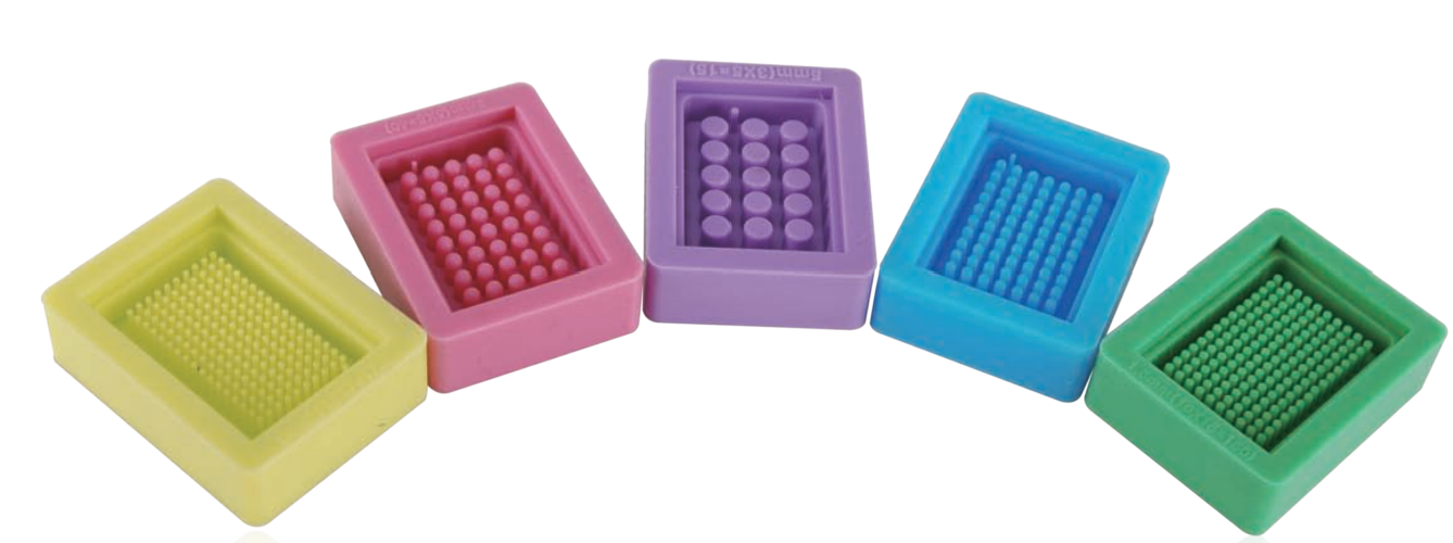
Core size : 3mm / 40 holes (5 x 8)
Core size : 1.5mm / 150 holes (10 x 15)
Core size : 5mm / 15 holes (3 x 5)
Core size : 2mm / 70 holes (7 x 10)
Recipient block mold kit is an alternative solution for the users to prepare the recipient block by themselves. The
recipient block can be used to excute TMA works with the manual tissue microarrayer called Quick Ray. Furthermore,
the researchers can prepare the hundreds of recipient blocks from a mold kit. For those who concerns over the price
of premade recipient blocks, Quick-Ray Mold Kit is an alternative solution. Made from silicon rubber, Quick-Ray
Mold kit allows users to build paraffin recipient blocks by themselves at a lower cost. The core sizes of each mold is
perfectly match with punchers of Quick-Ray. If stored and handed with care, hundreds of paraffin recipient blocks
can be produced by the Quick-Ray Mold Kit. But this recipient blocks can be applicable only for the manual tissue
microarrayer.
01Place the Recipient
block Mold kit in a dry
oven for 30 minutes at
70~80°C to warm-up
the mold kit.
04Dispense enough
liquid paraffin into the
embedding cassette
02Dispense liquid paraffin
(60~65°C) slowly into
the mold kit until the
top of core rods are
fully submerged.
05Solidify the embedding
cassette and the mold
kit at a normal room
temperature or at
about 4°C for 30~60
minutes.
If solidified at he lower
temperature, the block
may have cracks in it.
AParaffin dispense should
be done so slowly that
no bubbles are to be
formulated among the
core rods.
BWhen some bubbles are
formulated, remove them
with heated forceps.
06Separate the mold kit
from the embedding
cassette slowly and
carefully.
03Place an embedding
cassette on the mold
kit.
07Trim paraffin around
the periphery of the
recipient block.
Paraffin Block Trimmer
Quick Trim [UPBT-1011]
Paraffin block before trimming
Paraffin block after trimming
Specification
List Description
Power supply voltages 110 VAC / 220 VAC, 50/60 Hz
Weight (net) 47kg
Max Size (W x D x H) 580 x 477 x 280 mm
Operating temperature range Room temperature
Relative humidity Max. 80% non-condensing
Humidity during transportation/storage < 80%
Trimming capacity Max. 750 blocks per hour
Operating environment Indoor use only
Certification / Approval ISO / CE / FCC
The Paraffin Block Trimmer (generally called Wax
Trimmer, Paraffin Trimmer, Wax Remover, Paraffin
Remover, Dewaxer, etc) is one of the histology
equipments used for the histopathology. The Paraffin
Block Trimmer enables the researchers to trim the
paraffin blocks automatically.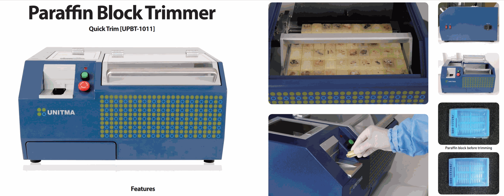
This instrument removes only paraffin residues
around the paraffin blocks without damage to the
tissue samples or the plastic cassette.
When users load the paraffin blocks on the inlet of the
instrument after the embedding process, the blocks
move into the trimming module to trim the paraffin
residues and the trimmed blocks are arrayed by 50
blocks in order in the array plate.
Eventually, this instrument also enables to reduce
the manually trimming time and to prevent the
users from any injuries caused by the conventional
trimming tools.
Features
• Save labor time of researchers
• Protect researchers from any physical damages
which may be caused by conventional trimming
tools.
• Max. 750 paraffin blocks can be trimmed in an hour.
• Semipermanant life time of trimming modules
• Accept the various kinds of the embedded blocks.
• 50 blocks can be trimmed at one time and continue
trimming by replacing the array plate with a new
one.
• The trimmed blocks are arrayed in order at the array
plate.